Decay indicator or caries indicator has been around for quite a while. I remember when I was taking histology in college and working in research labs at U.C. Berkeley, we used a cellular stain that reacted with the organic components of the cell. For several years in practice I wondered if certain stains could be used to identify cavities. A cavity is an area of demineralized enamel or dentin. Enamel and dentition are composed of a dense calcium rich matrix and relatively inorganic in its composition. The enamel is in fact one of the hardest naturally occurring substances allowing us to eat beer nuts and popcorn! When decay happens, bacteria penetrate into the tooth feeding on carbohydrates or sugars that we eat. The acid byproduct of the breakdown of these sugars demineralize the tooth structure. Once the decay has reached the dentin the bacteria can also feed on the existing tooth structure that is less mineralized and is softer. This bacteria and now much softer material can pick up organic stains more readily.
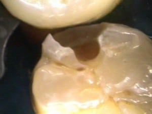
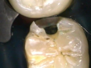
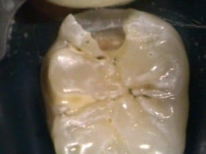
The other day I was removing the decay from this tooth. This cavity did not show up very well on X-rays and I used the Diagnodent laser to find it. I will go into the use of the Diagnodent later. As soon as I penetrated into this area with a small bur I dropped into a soft area, after some cleansing, I placed the decay indicator. Look how it stained the underlying area. This is remaining decay! This needs to be removed or the decay will remain under the filling. After cleaning and scraping the walls of the preparation the decay indicator is applied again. When there is no more uptake by decay indicator the cavity has been cleaned out and the tooth is ready for a filling. Without the decay indicator this cavity would not have been cleaned properly.

Before Decay Indicator

With Decay Indicator

After Decay Removal

