
The Difference a Millimeter Can Make
Correcting a Pathologic Joint Position using Mandibular Torque and a Fixed Orthosis
Neuromuscular dentists are often criticized as dentists who just open the bite. What happens when we open the bite and maybe things don’t go exactly as planned? The patient still may be suffering from symptoms, restorative treatment gets delayed and the patient begins losing confidence in the dentist to solve their problem. What happens when the patient is your father!
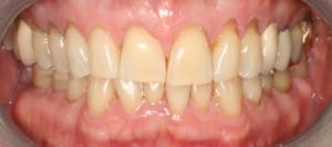
- Roy’s teeth before they were restored
My father, Roy Adler, who I treated for many years, had a severe traumatic painful episode in his left TMJ while eating an Italian ham sandwich on a French baguette. I have to be specific because my father takes his food very seriously. You see… I know his eating habits well. I also have a long dental history to fall back on, clues as to why this happened at this stage in his life. The clues that led directly to his episode helped me ultimately to determine the treatment that would relieve his pain… although the solution was not clear at first.
My father was well aware of my neuromuscular dental practice. He called from New York and told me he could no longer bring his teeth together normally. The pain was more dull than sharp. He experienced pain while chewing on both sides or tearing with his front teeth. The pain was strongest when first clamping down on food, but then lessened with subsequent chewing. He kept getting a dull ache when he pushed the jaw on the right side of the face to the left. Upon opening and closing the mouth he could hear bone rubbing against bone in the left TMJ. When he opened his mouth as wide as possible he sometimes felt the left TMJ catch, occasionally the right also. There was no pain or discomfort in the right joint. Opening the mouth wide to bite on food produced an ache, but doing the same maneuver without food did not. Directly after the initial event, there was a change in his bite. Upon waking in the morning he could close his teeth on the right side so that they met. However, soon after he would not be able to close down on the right side without difficulty, he would only hit on the left. So chewing food on the right side became a problem.
We used the Myotronics K-7 to evaluate his function. Scans showed that his muscles were firing at elevated levels in his temporal (temples) and cervical (neck) group. After 60 minutes of TENS (neuromuscular stimulation) there was improvement but the left cervical group remained elevated. His clench scores remained low, meaning that his muscles were not able to produce the proper amount of force when biting down. His bite was unstable. He had difficulty opening and was able to force himself to open to approximately 37 mm. The Sonography (scan of his joint noises) showed grating and popping noises on both sides. He was placed in a removable orthotic to open up his bite. The I-CAT scan without the orthotic shows boney degeneration.
When I placed my father in a neuromuscular removable orthotic there was little improvement. In fact the pain might have been slightly worse in the left TMJ although he was now in the proper bite. The orthotic had brought him down and forward, but his I-CAT scan revealed a disturbing fact. His joint on the left side was grinding against bone. Could we find a bite position that would relieve his pain within his joint? My father never really complained of muscular pain throughout this experience.
Repeated evaluations with the K-7 system showed that the orthotic was on Myo-trajectory – placing him in the correct bite. This created a dilemma; every Myo-bite taken left him in a bone to bone situation on the left side. We originally began with a removable orthotic to attempt to alleviate his condition. However his bite relationship without the orthotic did not leave him with a functional bite to eat with. We were not making much progress with the removable orthotic anyway. We made the decision to change to a fixed orthotic. We then used a new Myo-bite for Roy’s fixed orthotic. As you can see there were extensive degenerative changes now in the left joint as time went on. We can see the joint grinding against bone and the formation of joint mice or calcified bodies and bone fragments floating within the joint space. His opening became even more limited.
It was getting very hard to believe that things were going to get better. Was surgery going to be the only option? Was my father going to have to live with pain and suffering after paying for my education? He was traveling from New York to Colorado once a month for treatment. My father is a research mathematician. His scientific nature leads him to question everything in the minutest detail. He was well aware of my neuromuscular practice and successes with others. The problem had really hit home. The pressure was building. It was time to prove that neuromuscular dentistry was the way to go and was not quackery.
When we looked at his original radiograph things almost looked better before treatment. I had several thoughts about what to do. Should I remove the orthotic and see what happens. Well without the orthotic he was in constant pain with no bite. And he was in pain with the orthotic on Myo-centric trajectory but at least he could chew. I began to think about his dental history, the chronic break down of the left posterior teeth and subsequent crowning one after the other over the years.
The intra-oral signs were there long before his symptoms. There was a loss of vertical dimension (over closure of the bite), occlusal wear of anterior teeth and abfractions, tori (boney growths in his mouth), fractured teeth and deep bite. My father explained that he had chewed ice for years. As a teenager when his wisdom teeth were removed he began chewing almost exclusively on the left side after pain on the right. He continued this habit throughout his life until restored in a neuromuscular bite. The fact that we crowned almost the entire left side as these teeth fractured over the years was interesting. After years of restoring the left side he began fracturing the right side as well. I began placing crowns on the right side, one tooth after another. In August 2006 my father began complaining of pain in tooth number 31. There was a distal fracture extending through the pupil floor to the mesial. I placed a crown on number 31 and sent him home to NY. Shortly after the pain worsened and an endodontist determined the tooth was fractured through the root system. He removed the tooth and placed a bone graft. We placed an implant in the extraction site in August 2007. Now my father had lost his posterior stop on the right side, setting him up for catastrophic failure. One month after the implant was placed with a healing abutment at tissue height my father ate his infamous ham sandwich. The years of gradual loss of posterior vertical dimension first on one side then the other had finally taken their toll. As medical professionals we are told to “do no harm”. Well a whole lot of harm was done here over the years… one crown at a time.Perhaps the years of pathologic muscle function combined with the degeneration within his joints was preventing me from finding the ideal functional position. The atrophy of the system was not allowing TENS to correct the X/Y plane. If the torque created by the occlusal breakdown had led to this problem then maybe torquing the occlusion the opposite way might correct him.
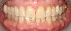
Roy’s New Pain Free Smile
On his next trip I decided to alter his orthotic. It seemed to make sense that we needed to torque the left side to rotate the condyle away from the bone. On his next trip we began with one hour of TENS. Then I began adding to the left side. I added a small amount of composite resin to tooth number 19 since it would give me the most secure stop. I checked my measurements and I had indeed increased the vertical on the left side by one millimeter. He was now hitting on both sides … Harder on the left but some on the right. I decided to leave him like that and let his own muscle function do the work. That night he noticed no difference in the pain he was experiencing while eating. I was lucky I could monitor the patient this way. I was able to watch every bite. He seemed like he was opening a little wider while eating. My father grabbed a raw carrot and crunched through it without complaining. I was afraid he was going to fracture the orthotic. The next day we tensed for one hour and began checking the bite. He was now hitting on both sides. With a slight adjustment to the right side and adding a little resin to the cusp tips of his right first bicuspid, he was now hitting evenly again.
My father returned to NY. I called him regularly for a week and asked how he was doing. My father could not tell me if he was improving. So I stopped calling. Two weeks later my father phoned… he realized he was completely out of pain. He has been out of pain ever since.
Three months later he flew out and we started with new I-CAT scans. The results were amazing. His left condyle had moved dramatically, the joint mice were gone. The K-7 scans showed great improvement in function. He was now able to open wide enough to restore him. He was on Myotrajectory. My father has had no recurrence of pain since adjusting his orthotic by correcting the torque. We then restored to the new position using the LVI protocol for full mouth reconstruction. My father remains pain free. What a difference one millimeter can make.
